Tổng hợp hệ vật liệu lai quang – từ định hướng cho ứng dụng nhiệt trị ung thư
524 lượt xemDOI:
https://doi.org/10.54939/1859-1043.j.mst.81.2022.128-137Từ khóa:
Coban ferit; Hạt nano bạc; Vật liệu lai hóa; Hiệu ứng sinh nhiệt; Hạt nano từ tính; Nhiệt trị ung thư.Tóm tắt
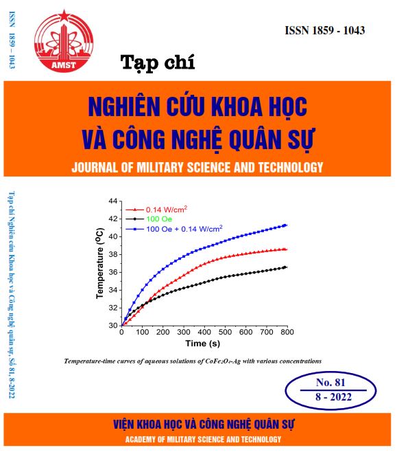
Các hạt nano từ tính sinh nhiệt dựa trên nền vật liệu CoFe2O4 là vật liệu tiềm năng cho hướng tiếp cận điều trị ung thư không xâm lấn. Tuy vậy, các hạt nano từ tính CoFe2O4 thường cho hiệu suất truyền nhiệt thấp, đã hạn chế nhiều trong phát triển các ứng dụng y sinh trong thực tế. Do đó, việc nghiên cứu phát triển các hệ vật liệu lai trên nền vật liệu từ cho hiệu suất truyền nhiệt cao hơn là cần thiết. Nghiên cứu này tập trung phát triển phương pháp đơn giản để tổng hợp trực tiếp hệ vật liệu lai quang từ coban ferrit (CoFe2O4) và hạt nano bạc (AgNPs), cho các ứng dụng yêu cầu hiệu suất sinh và truyền nhiệt cao. Nghiên cứu chỉ ra rằng, vật liệu lai quang-từ đã tổng hợp cho hiệu suất sinh nhiệt lớn hơn so với các vật liệu đơn lẻ (hạt nano bạc) và hạt nano từ CoFe2O4 khi cùng được kích thích dưới một điều kiện là từ trường xoay chiều hay laze. Kết quả thu được chứng minh rằng việc lai hóa giữa hạt nano từ và hạt nano bạc đã cho hiệu suất sinh nhiệt vượt trội. Vì vậy, hệ vật liệu tổng hợp CoFe2O4- là vật liệu tiềm năng cho lĩnh vực y sinh, điều trị ung thư bằng phương pháp không xâm lấn.
Tài liệu tham khảo
[1]. C.-H. Wu, J. Cook, S. Emelianov, and K. Sokolov, “Multimodal Magneto-Plasmonic Nanoclusters for Biomedical Applications,” Adv. Funct. Mater., vol. 24, no. 43, pp. 6862–6871, (2014). DOI: https://doi.org/10.1002/adfm.201401806
[2]. J. Kolosnjaj-Tabi, I. Marangon, A. Nicolas-Boluda, A. K. A. Silva, and F. Gazeau, “Nanoparticle-based hyperthermia, a local treatment modulating the tumor extracellular matrix,” Pharmacol. Res., vol. 126, pp. 123–137, (2017). DOI: https://doi.org/10.1016/j.phrs.2017.07.010
[3]. C. S. S. R. Kumar and F. Mohammad, “Magnetic nanomaterials for hyperthermia-based therapy and controlled drug delivery,” Adv. Drug Deliv. Rev., vol. 63, no. 9, pp. 789–808, (2011). DOI: https://doi.org/10.1016/j.addr.2011.03.008
[4]. P. H. Nam et al., “Polymer-coated cobalt ferrite nanoparticles: synthesis, characterization, and toxicity for hyperthermia applications,” New J. Chem., vol. 42, no. 17, pp. 14530–14541, (2018). DOI: https://doi.org/10.1039/C8NJ01701H
[5]. J.-H. Lee et al., “Exchange-coupled magnetic nanoparticles for efficient heat induction,” Nat. Nanotechnol., vol. 6, no. 7, pp. 418–422, (2011). DOI: https://doi.org/10.1038/nnano.2011.95
[6]. P. Kaur, M. L. Aliru, A. S. Chadha, A. Asea, and S. Krishnan, “Hyperthermia using nanoparticles – Promises and pitfalls,” Int. J. Hyperth., vol. 32, no. 1, pp. 76–88, (2016). DOI: https://doi.org/10.3109/02656736.2015.1120889
[7]. A. Espinosa, R. Di Corato, J. Kolosnjaj-Tabi, P. Flaud, T. Pellegrino, and C. Wilhelm, “Duality of Iron Oxide Nanoparticles in Cancer Therapy: Amplification of Heating Efficiency by Magnetic Hyperthermia and Photothermal Bimodal Treatment,” ACS Nano, vol. 10, no. 2, pp. 2436–2446, (2016).
[8]. M. Abdulla-Al-Mamun, Y. Kusumoto, T. Zannat, Y. Horie, and H. Manaka, “Au-ultrathin functionalized core–shell (Fe3O4@Au) monodispersed nanocubes for a combination of magnetic/plasmonic photothermal cancer cell killing,” RSC Adv., vol. 3, no. 21, p. 7816, (2013). DOI: https://doi.org/10.1039/c3ra21479f
[9]. A. Riedinger et al., “Subnanometer Local Temperature Probing and Remotely Controlled Drug Release Based on Azo-Functionalized Iron Oxide Nanoparticles,” Nano Lett., vol. 13, no. 6, pp. 2399–2406, (2013). DOI: https://doi.org/10.1021/nl400188q
[10]. M. F. Zarabi, N. Arshadi, A. Farhangi, and A. Akbarzadeh, “Preparation and Characterization of Gold Nanoparticles with Amino Acids, Examination of Their Stability,” Indian J. Clin. Biochem., vol. 29, no. 3, pp. 306–314, (2014). DOI: https://doi.org/10.1007/s12291-013-0358-4
[11]. W.-Z. Shen, S. Cetinel, K. Sharma, E. R. Borujeny, and C. Montemagno, “Peptide-functionalized iron oxide magnetic nanoparticle for gold mining,” J. Nanoparticle Res., vol. 19, no. 2, p. 74, (2017). DOI: https://doi.org/10.1007/s11051-017-3752-7
[12]. M. E. de Sousa et al., “Stability and Relaxation Mechanisms of Citric Acid Coated Magnetite Nanoparticles for Magnetic Hyperthermia,” J. Phys. Chem. C, vol. 117, no. 10, pp. 5436–5445, (2013). DOI: https://doi.org/10.1021/jp311556b
[13]. “Methods for Assessing Surface Cleanliness,” in Developments in Surface Contamination and Cleaning, Volume 12, Elsevier, pp. 23–105, (2019). DOI: https://doi.org/10.1016/B978-0-12-816081-7.00003-6
[14]. S. Campelj, D. Makovec, and M. Drofenik, “Preparation and properties of water-based magnetic fluids,” J. Phys. Condens. Matter, vol. 20, no. 20, p. 204101, (2008). DOI: https://doi.org/10.1088/0953-8984/20/20/204101
[15]. Q. Ding et al., “Shape-controlled fabrication of magnetite silver hybrid nanoparticles with high performance magnetic hyperthermia,” Biomaterials, vol. 124, pp. 35–46, (2017). DOI: https://doi.org/10.1016/j.biomaterials.2017.01.043
[16]. S. B. Waje, M. Hashim, W. D. W. Yusoff, and Z. Abbas, “X-ray diffraction studies on crystallite size evolution of CoFe2O4 nanoparticles prepared using mechanical alloying and sintering,” Appl. Surf. Sci., vol. 256, no. 10, pp. 3122–3127, (2010). DOI: https://doi.org/10.1016/j.apsusc.2009.11.084
[17]. S. Peng, C. Lei, Y. Ren, R. E. Cook, and Y. Sun, “Plasmonic/Magnetic Bifunctional Nanoparticles,” Angew. Chemie Int. Ed., vol. 50, no. 14, pp. 3158–3163, (2011). DOI: https://doi.org/10.1002/anie.201007794
[18]. Espinosa A., R. Di Corato, J. Kolosnjaj-Tabi, P. Flaud, T. Pellegrino, and C. Wilhelm, "Duality of iron oxide nanoparticles in cancer therapy: amplification of heating efficiency by magnetic hyperthermia and photothermal bimodal treatment" ACS Nano, vol. 10, pp. 2436-2446, (2016). DOI: https://doi.org/10.1021/acsnano.5b07249
[19]. Das R., N. Rinaldi-Montes, J. Alonso, Z. Amghouz, E. Garaio, J. A. Garcia, P. Gorria, J. A. Blanco, M.-H. Phan, and H. Srikanth, "Boosted hyperthermia therapy by combined AC magnetic and photothermal exposures in Ag/Fe3O4 nanoflowers" ACS Appl. Mater. Interfaces, vol. 8, pp. 25162-25169, (2016). DOI: https://doi.org/10.1021/acsami.6b09942